
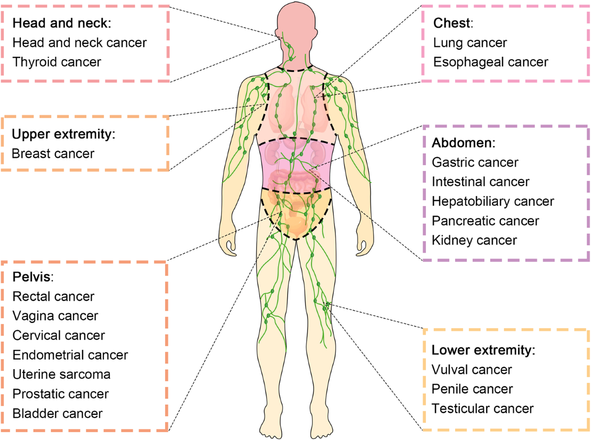
Lymph Nodes in Cancer: Understanding Their Role, Meaning, Spread, Staging, Diagnosis, and Treatment
Lymph nodes serve as vital components of the body’s immune system and lymphatic network. Functioning as biological filters, these structures continuously monitor and screen lymph fluid for pathogens, abnormal cells, and foreign bodies. Because malignant cells can utilize the lymphatic network as a pathway for dissemination, lymph nodes play a critical role in cancer detection, clinical staging, and the formulation of therapeutic strategies.
When healthcare providers announce that a malignancy has involved the regional lymph nodes, it generally signifies that malignant cells originating in a primary tumor have migrated into one or more nearby nodes. This scenario is fundamentally distinct from lymphoma, which is a primary malignancy arising directly from the lymphatic tissue and lymphocytes.
The detection of malignancy within a node does not inherently mean the disease has disseminated throughout the entire body. Instead, lymph node involvement is evaluated within the context of the specific cancer type, the total number and anatomical location of affected nodes, the physical size of the cancerous deposits, and the presence or absence of distant metastasis.
1. What Are Lymph Nodes?
A lymph node is a small, typically bean-shaped organ belonging to the lymphatic system. Hundreds of these nodes are distributed throughout the human body, organized in distinct clusters within regions such as:
-
The neck (cervical nodes)
-
The armpits (axillary nodes)
-
The chest (mediastinal and hilar nodes)
-
The abdomen and pelvis
-
The groin (inguinal nodes)
-
Surrounding major blood vessels and visceral organs
Interconnecting lymphatic vessels link these nodes, transporting a clear liquid known as lymph. This fluid consists of water, dissolved proteins, and various immune cells, prominently including lymphocytes.
The lymphatic system maintains vital fluid homeostasis, transports specific biological molecules, and provides critical immune defense. Inside each node, specialized immune cells inspect the passing fluid. When an infection arises, these immune cells become activated and proliferate, frequently causing the node to swell.
2. What Is the Relationship Between Lymph Nodes and Cancer?
Malignancy can involve the lymphatic network in two completely distinct manners:
Cancer Originating in the Lymphatic System
Certain malignancies begin directly within lymphatic structures. The primary examples include:
-
Hodgkin lymphoma
-
Non-Hodgkin lymphoma
These conditions are broadly classified under lymphomas, representing cancers of the blood and lymphatic tissues.
Cancer Spreading to Lymph Nodes
A much more common occurrence in solid tumors is secondary spread, where a cancer originates in a specific organ and subsequently travels to adjacent regional nodes. Examples include:
-
Breast malignancies spreading to axillary nodes in the armpit.
-
Oral, head, and neck cancers migrating to cervical nodes in the neck.
-
Lung carcinomas involving nodes located within the chest cavity.
-
Colorectal tumors metastasizing to nodes surrounding the intestinal tract.
-
Melanoma spreading to regional skin-draining nodes.
The specific nodes affected depend entirely on the primary tumor’s site and the natural drainage pathways of the local lymphatic architecture. Crucially, when secondary cancer cells are identified inside a lymph node, they retain the classification of the primary tumor. For instance, breast cancer cells discovered in an axillary node are treated and diagnosed as metastatic breast cancer, not as lymphoma.
3. How Does Cancer Reach a Lymph Node?
In early stages, malignant cells remain confined to their primary tumor site. However, as the neoplasm progresses, certain cells acquire the capacity to invade adjacent healthy tissue boundaries.
Some of these cells can penetrate the walls of nearby lymphatic vessels. Once inside, they are swept along with the flowing lymph toward regional nodes. This sequence can be summarized as follows:
$$\text{Primary Tumor} \longrightarrow \text{Lymphatic Vessel} \longrightarrow \text{Regional Lymph Node} \longrightarrow \text{Potential Further Spread}$$
Not every malignant cell entering a lymphatic vessel successfully establishes a secondary colony; many perish or are eradicated by immune defenses. Nevertheless, aggressive cells may survive, settle, and multiply within the node. Additionally, dissemination can occur via the bloodstream, making both the lymphatic and vascular systems primary highways for cancer migration.
4. Why Are Lymph Nodes Crucial in Cancer Staging?
Examining lymph nodes provides indispensable data for staging a cancer. Staging classifies the extent and severity of the disease at a given time, guiding clinical decision-making.
The globally accepted standard is the TNM staging system:
-
T (Tumor): Denotes the dimensions and/or local tissue invasion of the primary mass.
-
N (Nodes): Describes whether regional lymph nodes contain cancer, incorporating their quantity, distribution, and extent of involvement.
-
M (Metastasis): Indicates whether the cancer has spread to distant anatomical sites or organs.
For example, a staging designation of T2 N1 M0 specifies particular primary tumor characteristics, regional node involvement, and an absence of detected distant spread. The exact parameters for values like N1 vary widely depending on the underlying cancer type. Generally:
-
N0: No regional node involvement identified.
-
N1, N2, N3: Increasing levels of regional node involvement, defined specifically by individual cancer protocols.
-
M0: No distant metastasis detected.
-
M1: Distant metastasis confirmed.
Consequently, simply knowing a node is “positive” does not reveal the complete stage of the disease.
5. Does a Swollen Lymph Node Always Indicate Cancer?
No. This is a critical distinction.
Lymph nodes frequently enlarge because the immune system is actively mounting a defense against:
-
Viral infections (e.g., common cold, influenza)
-
Bacterial infections (e.g., strep throat)
-
Localized inflammation
-
Non-malignant immune reactions
For instance, neck nodes commonly swell during upper respiratory infections. While malignancies can also trigger nodal enlargement, physical examinations alone cannot reliably differentiate between benign infections and neoplastic involvement. Furthermore, a node packed with malignant cells may occasionally remain normal in size and undetectable by touch, whereas an enlarged node may be entirely benign. This uncertainty necessitates imaging or tissue biopsy when clinical suspicion arises.
6. What Do Malignant Lymph Nodes Look Like?
No single physical appearance universally proves that a node harbors cancer. Clinicians look for warning signs during physical checks and imaging studies, such as persistent enlargement, irregular or abnormal morphology, or a hard, fixed texture.
However, imaging alone cannot always establish a definitive diagnosis, as a normal-appearing node can still harbor microscopic deposits of cancer cells. For absolute confirmation, microscopic evaluation by a pathologist via biopsy is required.
7. How Are Lymph Nodes Evaluated?
Healthcare providers utilize several diagnostic modalities to investigate lymph nodes:
-
Physical Examination: Palpable superficial nodes in the neck, armpits, and groin can be evaluated for size, consistency, mobility, and tenderness, though physical findings alone are not definitive.
-
Ultrasound: Highly effective for superficial nodes, providing clear visualization of internal architecture to help determine if a biopsy is warranted.
-
CT Scan: Computed tomography scans assess deep nodes within the chest, abdomen, or pelvis, serving as a pillar of cancer staging.
-
MRI: Magnetic resonance imaging offers detailed soft-tissue resolution, valuable for specialized anatomical regions.
-
PET/CT: Combines metabolic tracking with anatomical imaging to spot areas of high cellular activity, though active inflammation can occasionally mimic malignant findings.
8. What Is a Lymph Node Biopsy?
A biopsy involves extracting cells or tissue from a node for microscopic analysis. Techniques include:
-
Fine-Needle Aspiration (FNA): Uses a thin needle to withdraw cells.
-
Core-Needle Biopsy: Uses a larger needle to secure a small cylinder of tissue.
-
Excisional Biopsy: Surgical removal of an entire lymph node.
-
Sentinel Lymph Node Biopsy: Removal of the first filtering nodes in a drainage basin.
Pathologists analyze these samples to confirm the presence of cancer, detail the burden of disease, and report specific microscopic characteristics.
9. What Is a Sentinel Lymph Node?
The sentinel lymph node represents the initial node or group of nodes reached by lymphatic fluid draining directly from a primary tumor. This concept is fundamental in managing conditions like breast cancer and melanoma.
During a sentinel lymph node biopsy, surgeons inject a safe tracer and/or a blue dye near the tumor to trace its drainage pathway. The identified sentinel nodes are then removed and analyzed. If these primary nodes are clear of cancer, extensive removal of subsequent regional nodes may be safely avoided, minimizing patient morbidity.
10. What Does a “Positive Lymph Node” Signify?
A positive lymph node means that microscopic examination has identified malignant cells within that tissue. Conversely, a negative node contains no detected cancer.
If a surgeon extracts ten nodes and pathology discovers cancer in two of them, the report might read:
2 of 10 lymph nodes positive for metastatic carcinoma.
The clinical meaning of this finding depends entirely on the cancer type, the proportion of positive-to-examined nodes, the anatomical location, the size of the deposits, and whether the cancer has breached the outer capsule of the node.
11. Micrometastases and Isolated Tumor Cells
Malignancy within a node is not always a binary presence or absence. Pathologists can detect microscopic disease burdens categorized as:
-
Isolated Tumor Cells (ITCs): Extremely small clusters of cells.
-
Micrometastases: Tiny deposits (typically ranging between 0.2 mm and 2 mm).
-
Macrometastases: Larger, clinically apparent cancer deposits.
Thresholds and staging implications vary widely across different organ systems, requiring interpretation within specific disease guidelines.
12. Does Lymph Node Involvement Equal Stage IV Cancer?
Not necessarily. This represents one of the most common misconceptions regarding cancer staging.
Malignancy frequently spreads from a primary organ to regional, nearby nodes without advancing to distant organs. The National Cancer Institute classifies regional node involvement as localized or regional spread, whereas Stage IV typically denotes distant metastasis to remote organ systems.
Therefore, a positive regional lymph node does not automatically translate to Stage IV disease, though exact staging rules remain unique to each cancer type.
13. Why Is Lymph Node Involvement Clinically Significant?
Finding cancer within regional nodes signals that malignant cells have breached the boundaries of the primary tumor and entered the lymphatic system. This insight fundamentally alters therapeutic planning.
Depending on the specific cancer diagnosis, a positive node finding may prompt multidisciplinary teams to consider additional treatments, such as:
-
Surgical resection
-
Radiation therapy
-
Systemic chemotherapy
-
Hormone therapy
-
Targeted molecular therapies
-
Immunotherapy
14. What Is Lymph Node Dissection?
A lymph node dissection (or lymphadenectomy) is a surgical intervention where multiple nodes are extracted from a specific anatomical region. This procedure is performed to achieve accurate pathological staging, halt regional disease progression, or treat confirmed nodal spread.
While removing nodes does not compromise the body’s entire immune network—given its vast and redundant distribution—it can lead to side effects, most notably lymphedema.
15. What Is Lymphedema?
Lymphedema is chronic tissue swelling caused by the accumulation of protein-rich lymphatic fluid, typically triggered by the surgical removal of nodes or radiation damage to lymphatic channels. For example, axillary node surgery for breast cancer can result in arm swelling, while pelvic or groin dissections can cause leg swelling. Symptoms involve persistent swelling, a sensation of heaviness, reduced limb mobility, and potential skin texture changes. Although often manageable, lymphedema can become a lifelong condition.
16. Can Cancer Spread Beyond the Lymph Nodes?
Yes. Malignancy can progress from a primary site to regional nodes and subsequently disseminate via the vascular or lymphatic systems to distant organs. However, regional nodal involvement does not guarantee that distant metastasis has occurred. Comprehensive clinical workups utilizing advanced imaging and laboratory tests are required to determine whether disease is localized, regionally advanced, or metastatic.
17. Lymph Nodes Across Different Cancer Types
The diagnostic and prognostic weight of lymph node evaluation varies significantly depending on the malignancy:
-
Breast Cancer: Axillary nodes are critical indicators; sentinel biopsies guide adjuvant treatment.
-
Lung Cancer: Mediastinal and chest nodes dictate staging and operability.
-
Head and Neck Cancers: Cervical nodes in the neck require careful mapping and evaluation.
-
Colorectal Cancer: Regional intestinal nodes are routinely harvested during bowel resections to determine pathological stage.
-
Melanoma: Sentinel node status dictates the necessity of systemic or secondary therapies.
-
Prostate Cancer: Pelvic nodes influence risk stratification and treatment intensity.
18. Interpreting a Lymph Node Pathology Report
A comprehensive pathology report details vital data points, including:
-
The total number of nodes evaluated.
-
The exact number of nodes harboring malignant cells.
-
The specific histological subtype of the cancer.
-
The physical dimensions of any metastatic deposits.
-
Whether cancerous growth extends beyond the outer capsule of the node into surrounding soft tissue.
19. Summary: When to Seek Medical Evaluation
While the vast majority of swollen lymph nodes are benign responses to common viral or bacterial infections, persistent or concerning changes warrant professional medical assessment. Individuals should schedule an evaluation if they notice:
-
Swelling that persists without a clear explanation or continues to enlarge.
-
Nodes that feel unusually hard, rubbery, or fixed in place.
-
Multiple unexplained enlarged nodes.
-
Systemic symptoms such as unexplained weight loss, persistent fevers, drenching night sweats, or profound fatigue.
10 Facts That Explain Why Data Is the New Global Currency | Maya
Medical Disclaimer: This article is for general educational purposes only and does not constitute medical advice, diagnosis, or treatment. Cancer and lymph-node involvement vary from person to person. Always consult a qualified healthcare professional for proper evaluation and personalized medical guidance.